1. Theoretical Foundations: Sleep as an Active Computational State
For centuries, sleep was conceptualized as a passive, dormant state—a mere cessation of consciousness necessitated by physical exhaustion. Modern clinical neuroscience and neuroimaging have completely overturned this dogma: sleep is now recognized as an intensely active, highly coordinated metabolic and computational state essential for human intellectual function. During nocturnal slumber, the brain consumes almost as much ATP as during active waking hours, reallocating its massive bioenergetic budget from sensory perception and motor action toward structural macromolecular repair, synaptic recalibration, and long-term memory consolidation.
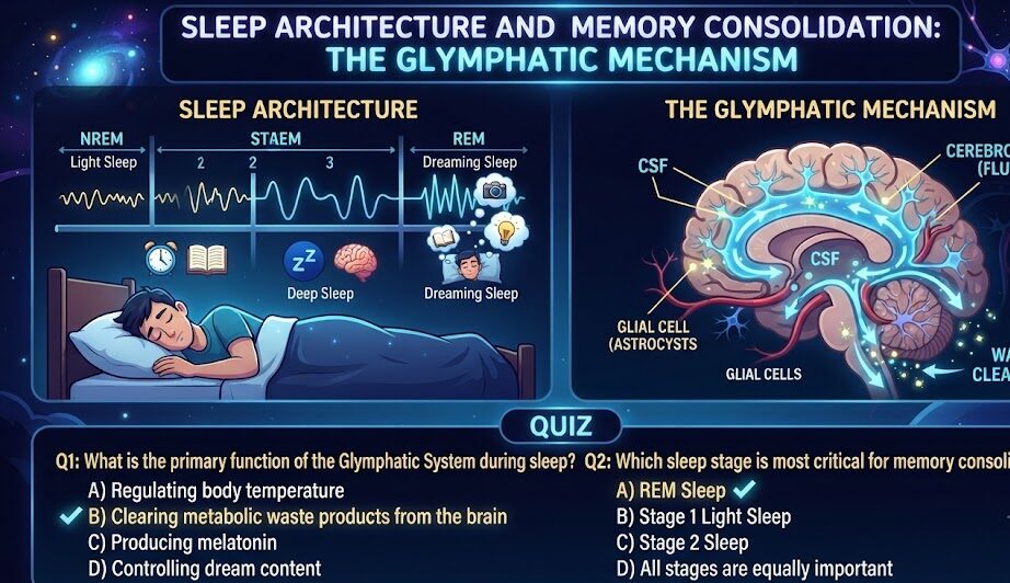
The architectural organization of human sleep is partitioned into distinct ultradian cycles of non-rapid eye movement (NREM) sleep—further stratified into stages N1, N2, and N3 (slow-wave sleep)—and rapid eye movement (REM) sleep. Each cycle persists for approximately 90 to 110 minutes, recurring 4 to 6 times per night. Rather than functioning redundantly, each specific stage executes non-overlapping computational algorithms: NREM slow-wave sleep coordinates the systemic transfer of episodic memories from temporary hippocampal stores to stable neocortical networks, while REM sleep integrates disparate memories into abstract associational schemas, fueling creative problem-solving and emotional equilibrium.
Crucially, sleep serves an indispensable maintenance function through the recently characterized glymphatic system. Discovered by Dr. Maiken Nedergaard and colleagues, this glial-dependent waste clearance pathway utilizes convective cerebrospinal fluid (CSF) influx to flush neurotoxic metabolic byproducts—including amyloid-beta, hyperphosphorylated tau, and alpha-synuclein—from interstitial space. Without sufficient, architecturally sound slow-wave sleep, the brain suffers chronic toxic metabolite stagnation, precipitating progressive executive degradation and accelerated neurodegeneration.
2. Neurobiological Mechanics of Memory Consolidation: The Tripartite Dialogue
The transformation of labile, newly acquired experiences into permanent long-term memory relies upon a precisely synchronized neurophysiological process known as the two-stage memory model, mediated by a tripartite dialogue during NREM Stage 3 sleep:
1. Neocortical Slow Oscillations (<1 Hz): Originating within the prefrontal cortex, these high-amplitude, low-frequency electrical waves propagate anteroposteriorly across the neocortex. They establish widespread, synchronized windows of neuronal excitability (up-states) and silence (down-states), orchestrating global cortical communication.
2. Thalamocortical Sleep Spindles (11–16 Hz): Triggered during the up-states of cortical slow oscillations, bursts of rhythmic sinusoidal activity are generated by the thalamic reticular nucleus. Spindles project to the cortex, triggering a massive influx of intracellular calcium into postsynaptic pyramidal dendrites, which activates CaMKII and primes synapses for persistent structural plasticity.
3. Hippocampal Sharp-Wave Ripples (150–250 Hz): Nestled precisely within the troughs of sleep spindles, the hippocampal CA3 and CA1 regions fire high-frequency sharp-wave ripples. During these ripples, neural ensembles that encoded waking experiences replay in accelerated time (up to 20 times faster than waking speed). This replay drives the transfer of information from the temporary, high-turnover hippocampal buffer into distributed neocortical sites for permanent storage.
4. The Synaptic Homeostasis Hypothesis (SHY): Pioneered by Giulio Tononi and Chiara Cirelli, the Synaptic Homeostasis Hypothesis posits that wakefulness is accompanied by net synaptic potentiation across widespread cortical circuits as the brain continually learns and adapts. This progressive increase in synaptic strength places unsustainable demands on cellular space, energy, and supply of neurotrophic factors. During slow-wave sleep, non-specific global synaptic downscaling occurs: weaker, task-irrelevant synaptic connections are systematically pruned and Depotentiated, while biologically salient, spindle-reinforced connections are spared. This computational renormalization restores the signal-to-noise ratio of the cerebral cortex, freeing up fresh bioenergetic capacity for new learning on subsequent days.
3. The Glymphatic System and Aquaporin-4 (AQP4) Dynamics
During waking consciousness, the dense, continuous firing of neurons requires a tightly packed interstitial matrix. However, this spatial density severely restricts the diffusive clearance of large macromolecular solutes. The glymphatic system solves this biological bottleneck via a specialized convective fluid transport mechanism:
Astrocytic Endfeet and AQP4 Water Channels: Cerebral blood vessels are enveloped by vascular endfeet of specialized astrocytes expressing exceptionally high densities of aquaporin-4 (AQP4) water channels. Cerebrospinal fluid (CSF) from the subarachnoid space enters the periarterial (Virchow-Robin) spaces surrounding cerebral arteries, driven by arterial pulsatility generated by cardiac contractions.
The 60% Interstitial Volume Expansion: In foundational in vivo two-photon imaging experiments, Nedergaard et al. demonstrated that upon transitioning into slow-wave sleep, prefrontal and cortical interstitial space volume expands by an astonishing 60%. This dramatic expansion is driven by a steep decline in central locus coeruleus norepinephrine release. With interstitial resistance eliminated, CSF rushes through AQP4 channels into the brain parenchyma, mixing with interstitial fluid (ISF) and stripping neurotoxic solutes toward perivenous spaces for drainage into deep cervical lymph nodes.
When sleep is fragmented or shortened, this convective hydraulic cleansing process is prematurely truncated. Un-cleared metabolic debris accumulates within the interstitial matrix, creating diffuse micro-inflammation that impairs frontoparietal synaptic transmission within less than 24 hours.
4. REM Sleep, Emotional Homeostasis, and Associational Creativity
While NREM slow-wave sleep is the master of declarative fact consolidation and metabolic cleansing, REM sleep serves as the brain’s nocturnal emotional therapist and creative incubator:
The Neurochemistry of REM: High Acetylcholine, Zero Noradrenaline: During REM sleep, the central brain enters a unique neurochemical state found at no other time in life: acetylcholine levels surge to concentrations higher than during wakefulness, promoting intense cortical plasticity, while locus coeruleus noradrenaline release ceases completely. This enables the brain to reactivate emotionally charged memories without triggering the visceral, sympathetic autonomic stress responses that accompanied the original event. Over repeated REM cycles, this process “strips the emotional charge” from difficult memories, preserving the factual lesson while restoring emotional stability.
Cross-Domain Semantic Associational Linking: Functional neuroimaging during REM demonstrates high functional connectivity across the anterior cingulate, temporoparietal junctions, and limbic structures, while the lateral prefrontal cortex remains largely quiescent. Unconstrained by rigid executive rules, the sleeping brain creates novel, non-linear associational connections across disparate semantic databases. This explains why complex mathematical breakthroughs, linguistic epiphanies, and creative insights frequently emerge immediately upon waking from deep, undisturbed sleep.
5. Molecular Biomarkers of Sleep Architecture Dysfunction
Chronic sleep disruption manifests as measurable neurobiological and physiological perturbations:
Elevated CSF/Plasma Biomarkers: Positron emission tomography (PET) and cerebrospinal fluid assays reveal that even one night of acute total sleep deprivation triggers an immediate 25% to 30% surge in measurable amyloid-beta 42 and phosphorylated tau (p-tau181) levels in human subjects. These elevations directly parallel transient decrements in spatial navigational accuracy and verbal memory retrieval.
Neuroinflammatory Cytokine Cascade: Fragmented slow-wave sleep stimulates microglial activation and systemic elevations of tumor necrosis factor-alpha (TNF-alpha), interleukin-6 (IL-6), and high-sensitivity C-reactive protein (hs-CRP). This chronic low-grade neuroinflammation suppresses hippocampal adult neurogenesis in the subgranular zone of the dentate gyrus and degrades prefrontal white matter tract microstructure over time.
Comparative Neuroanalytical Framework
To quantify the physiological, metabolic, and behavioral divergence across attentional states, the following high-density comparative matrix contrasts baseline operations against acute focus trajectories:
| Sleep Architecture Stage | Dominant EEG Frequency Band | Primary Neurochemical Profile | Core Cognitive & Metabolic Function |
|---|---|---|---|
| NREM Stage 2 (Light Sleep) | Theta (4–8 Hz) + Sleep Spindles (11–16 Hz) | Moderate GABA; declining monoamines | Motor procedural memory; calcium influx into cortical spines |
| NREM Stage 3 (Slow-Wave Sleep) | Delta (0.5–4 Hz) + Slow Oscillations (<1 Hz) | Minimal norepinephrine; high growth hormone | Hippocampal-to-cortical transfer; 60% glymphatic clearance surge; SHY synaptic renormalization |
| REM (Rapid Eye Movement) | Desynchronized beta/theta (sawtooth waves) | Maximal acetylcholine; zero noradrenaline | Emotional affective recalibration; cross-domain creative synthesis |
| Acute Sleep Deprivation State | Local cortical slow-wave intrusions during wakefulness | Adenosine accumulation; compensatory cortisol surge | Micro-sleeps; severe dlPFC decoupling; 400% elevated error rate |
Actionable Clinical & Cognitive Protocols
Translating neurobiological theory into measurable intellectual performance requires standardized behavioral frameworks designed to optimize synaptic signaling and preserve metabolic substrates:
Protocol A: Slow-Wave Thermal Priming
Slow-wave sleep architecture is directly modulated by ambient and core temperature dynamics. To maximize early-night slow-wave sleep percentage, maintain bedroom ambient temperature strictly between 18°C and 19.5°C (65°F–67°F). Take a warm shower or bath (40°C–42°C) 90 minutes prior to sleep; this triggers rapid peripheral vasodilation (heat dumping through the soles of feet and palms), facilitating the critical 1°C drop in core body temperature required for rapid delta sleep initiation.
Protocol B: Gastrointestinal Glymphatic Protection Window
Late-night food consumption forces the parasympathetic and circulatory system to sustain heavy splanchnic blood flow, preventing the normal drop in nocturnal heart rate and elevating core body temperature. Conclude all caloric intake at least 3 hours prior to sleep. This ensures that the cardiovascular system enters its nocturnal “dipping” phase, maximizing arterial pulsatility in cerebral vessels—the primary mechanical driver of convective glymphatic CSF fluid flow.
Protocol C: Lateral Decubitus Sleep Posture
In rodent and human neuroimaging research evaluating glymphatic clearance efficiency across anatomical sleeping positions, lateral decubitus (side-sleeping) positions demonstrated significantly superior CSF-ISF exchange compared to prone (stomach) or supine (back) positions. Lateral sleeping optimizes gravitational and hemodynamic drainage through the jugular and vertebral venous plexuses, accelerating the clearance of metabolic solutes from the cortex.
Common Neuromyths, Pitfalls & Diagnostic Misattributions
Pervasive clinical misconceptions surround sleep physiology:
- Sedation is Not Sleep: Consuming central nervous system depressants (such as alcohol, benzodiazepines, or Z-drugs) induces sedation by allosterically modulating GABA-A receptors, but violently disrupts natural sleep architecture. Alcohol severely suppresses REM sleep and fragments slow-wave sleep spindles, effectively eliminating memory consolidation despite 8 hours of unconsciousness.
- The Myth of Catch-Up Sleep: Sleeping 10 hours on Sunday cannot reverse the synaptic damage, cognitive degradation, or metabolic dysregulation incurred across five consecutive nights of 5-hour sleep. While subjective sleepiness may dissipate, frontoparietal connectivity and executive error rates remain clinically impaired.
- Ignoring Sleep Consistency: Variable bedtimes and wake times create constant circadian desynchrony. Going to bed at 22:00 one night and 02:00 the next disrupts the precise alignment between SCN-governed REM windows and homeostatic slow-wave sleep pressure, depriving the brain of critical consolidation phases.
Frequently Asked Clinical Questions (FAQ)
How does caffeine consumed in the afternoon impact nocturnal slow-wave sleep?
Caffeine possesses an average elimination half-life of 5 to 7 hours and a quarter-life of up to 12 hours. Even if an individual falls asleep easily, residual caffeine molecules occupying cerebral adenosine A1/A2A receptors decrease Stage 3 slow-wave sleep depth by up to 30%, severely blunting glymphatic clearance and declarative memory consolidation.
Can daytime naps replace nocturnal sleep for memory consolidation?
A 60-to-90 minute daytime nap that encompasses both slow-wave sleep and REM can provide measurable short-term benefits for memory consolidation and alertness. However, naps cannot fully replace nocturnal sleep, which is required to complete multiple synchronized cycles of glymphatic waste clearance and hormonal regulation.
Why do we forget most of our dreams upon waking?
During REM sleep, central norepinephrine release is completely shut off, and the hippocampus is primarily operating in an outward ‘playback’ mode rather than an inward ‘recording’ mode. Without norepinephrine to stabilize immediate synaptic plasticity, dream narratives dissipate within seconds unless immediately rehearsed upon waking.
How does sleep deprivation cause emotional instability?
fMRI research reveals that following a single night of sleep deprivation, functional connectivity between the prefrontal cortex (responsible for rational oversight) and the amygdala (emotional threat detection) decreases by over 60%. The amygdala becomes hyper-reactive, causing minor stressors to trigger disproportionate emotional reactions.
What are the early cognitive symptoms of glymphatic clearance failure?
Early symptoms include persistent morning brain fog lasting longer than 90 minutes, reduced short-term working memory retention, difficulty synthesizing novel conceptual connections, and increased reliance on high-dose stimulants to achieve baseline alertness.
Peer-Reviewed Scholarly References
- Nedergaard, M., & Goldman, S. A. (2020). Glymphatic failure as a final common pathway to dementia. Science, 370(6512), 50-56.
- Xie, L., et al. (2013). Sleep drives metabolite clearance from the adult brain. Science, 342(6156), 373-377.
- Diekelmann, S., & Born, J. (2010). The memory function of sleep. Nature Reviews Neuroscience, 11(2), 114-126.
- Walker, M. P., & Stickgold, R. (2006). Sleep, memory, and plasticity. Annual Review of Psychology, 57, 139-166.
- Rasch, B., & Born, J. (2013). About sleep’s role in memory. Physiological Reviews, 93(2), 681-766.
- Tononi, G., & Cirelli, C. (2014). Sleep and the price of plasticity: from synaptic and cellular homeostasis to memory consolidation and integration. Neuron, 81(1), 12-34.